Neurobiology - the visual sense studied in zebrafish
Vision is achieved thanks to possible by specialized cells in the eye, able to convert light into a biological signal that can be interpreted by the brain.
Photoreceptors
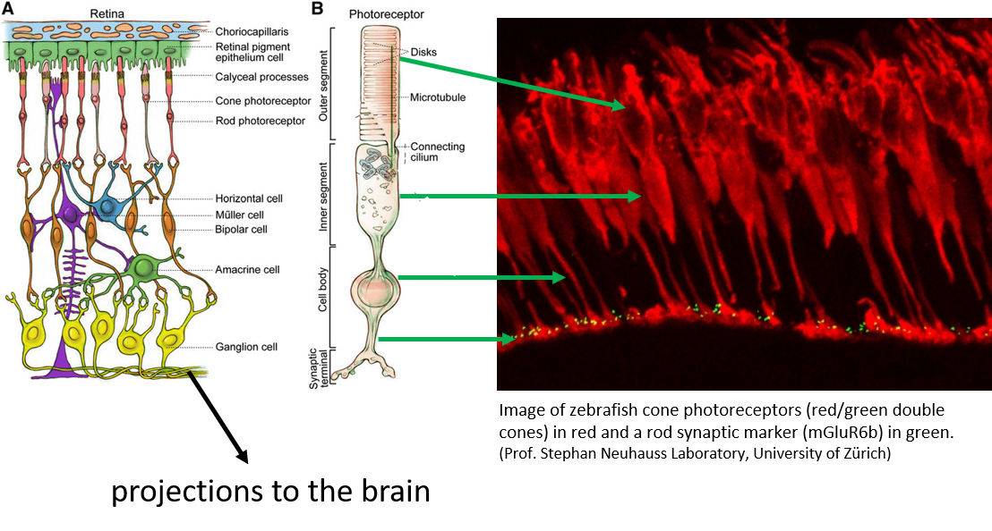
These cells are called photoreceptors and come in two shapes. Rod photoreceptors are highly light sensitive but cannot convey color information, while cone photoreceptor function under brighter light conditions and can convey not only brightness but also color information. There is only one rod phocoreceptor type in vertebrates, while there are up to four cone types that are tuned to cover different colors of the light spectrum. In humans, there are three subtypes that are most sensitive to blue, green and red light, while zebrafish have an additional subtype sensitive to ultraviolet light. Neuroscientists are fascinated by the vast range of light intensities that the visual system can handle. Our eyes support vision on a moonlit night as well as on a bright summer day. The brightest light is about a 100 billionth times brighter than the dimmest light that we can perceive (think of a one followed by 11 zeros, or the range of weight from 1 gram and 10 million tons!).
Studying visual senses in Zebrafish
This remarkable task is partially achieved by having a dedicated low light system (using rod photoreceptors) and a bright light system (using cone photoreceptors) and in part by sophisticated biochemical regulatory mechanisms intrinsic to photoreceptors. The latter system is especially important for cone photoreceptors, which have the largest range of light sensitivity and on which we humans rely on for most of our daily lifes (thanks to artificial lighting). The intriguing biology of cone photoreceptors is difficult to study in humans for practical and ethical reasons, but also nocturnal rodents such as mice are not well suited, due to their paucity of cone photoreceptors. This is where the characteristics of the zebrafish visual system come into play, as the cone photoreceptors dominate the zebrafish vision. In fact, young larvae rely exclusively on cone photoreceptor function to support vision. Others and we and others have developed a suite of behavioral and electrophysiological methods to precisely measure visual performance. As a result, we are able to detect even subtle visual defects at the larval stage. Encouragingly, electrophysiological recordings from the eye are directly comparable to measurements from mice and humans.
Within any cell, including a cone photoreceptor cell, lies the genetic material. The genetic material is the DNA that contains the blueprint for the composition of each cell type. Some proteins/molecules are produced only by a specific cell type. Proteins can act as messengers that affect other cells or processes within the cell and can also affect their function. Imagine a construction site: proteins are the workers while DNA is the project leader in the nucleus. Many of these proteins are also known to malfunction in inherited ocular diseases of humans. Other molecules from the outside can also influence cells. In all neurons, calcium is a messenger substance that triggers reactions. Sophisticated read-out measurements can now be applied to zebrafish larvae in which certain protein functions have been compromised by pharmacological or genetic means.
Studies have shown that adaptation of cone photoreceptors to light is dominated by a calcium-dependent regulatory mechanisms. Calcium influences the activation of visual pigments within a photoreceptor. Artificial proteins can mimic and influence natural proteins that are specific to cone photoreceptors. In addition, there is a greater diversity of calcium binding proteins in cones. A deeper understanding of these mechanisms will not only address the long-standing question of adaptation, but will also "shed light" on blinding diseases, such as retinal dystrophies and age-related macular degeneration.


